Quick Tips
Supraclavicular Block
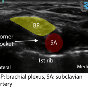
- Deposit local anesthetic within the ‘corner pocket’ to ensure reliable blockade of the inferior trunk, which gives rise to the ulnar nerve.
- The brachial plexus, subclavian artery and first rib form the borders of the corner pocket.
PECS Block
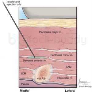
- Aim the needle towards the 4th rib when performing a PECS II block to minimize the risk of pneumothorax.
- The rib acts as a back stop to prevent the needle from advancing deep towards the pleura.
Anterior Quadratus Lumborum (QL3)
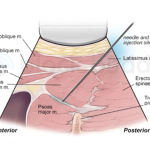
- Avoid depositing local anesthetic within the psoas muscle to minimize the risk of lumbar plexus blockade and subsequent lower extremity weakness.
- All of the major branches of the lumbar plexus pass through the psoas muscle.
Pericapsular Nerve Group (PENG) Block
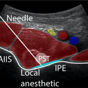
- After advancing the needle through the psoas tendon (PST) and making contact with the iliopubic eminence (IPE), rotate the needle hub between the index finger and thumb to ensure passage through the tendon.
- Confirm correct needle tip placement by observing the spread of local anesthetic along the pubic bone posterior to the psoas tendon.
iPACK Block
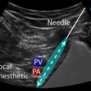
- Avoid advancing the needle tip more than 2cm beyond the lateral border of the popliteal artery to minimize common peroneal nerve blockade and foot drop.
Popliteal Sciatic Nerve Block
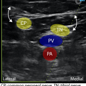
- Enhance identification of the tibial and common peroneal nerves on ultrasound by having the patient dorsi and plantar flex the foot.
- Plantar flexion: tibial nerve is elevated towards the transducer.
- Dorsiflexion: common peroneal is elevated towards the transducer.
Intermediate Cervical Plexus Block
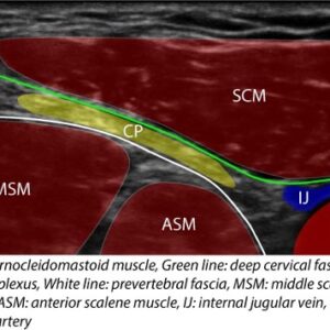
- During an intermediate cervical plexus block, avoid advancing the needle tip through the prevertebral fascia to avoid inadvertent phrenic nerve blockade.
- The prevertebral fascia passes over top of the scalene muscles and phrenic nerve.
Infraclavicular Block
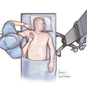
- Abducting the arm 90 degrees at the shoulder during an infraclavicular block may help reduce the depth of the brachial plexus and create more space between the transducer and clavicle for needle insertion.
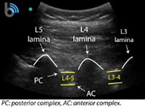
Neuraxial Scanning
- Using ultrasound for neuraxial block placement can be useful for identifying the correct intervertebral space, determining the correct angle for needle insertion, and measuring the ligamentum flavum/dura depth.
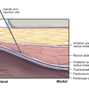
Rectus Sheath Block
- When performing a rectus sheath block, use a blunt tip needle for more tactile sensation and to reduce the risk of perforating the posterior wall of the rectus sheath.
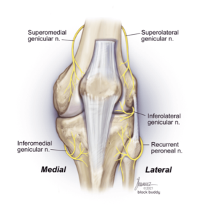
Genicular Nerve Block
- When blocking the genicular nerves for knee arthroplasty, the inferolateral genicular nerve is not usually targeted due to the proximity of the common peroneal nerve and the risk for foot drop.
Sciatic Nerve Block
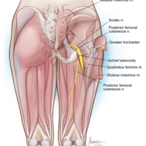
- The posterior femoral cutaneous nerve (PFCN) begins to divide away from the sciatic nerve near the gluteal crease.
- Targeting the sciatic nerve at this level may result in inadequate blockade of the PFCN.
